C57BL/6JNifdc-Cd4tm1(EGFP-DTR-luc)Bcgen/Bcgen • 113729
| Product name | B-Cd4-EGFP-DTR-Luc mice |
|---|---|
| Catalog number | 113729 |
| Strain name | C57BL/6JNifdc-Cd4tm1(EGFP-DTR-luc)Bcgen/Bcgen |
| Strain background | C57BL/6JNifdc |
| NCBI gene ID | 12504 (Mouse) |
| Aliases | L3T4; Ly-4 |
Gene targeting strategy for B-Cd4-EGFP-DTR-Luc mice. A construct composed of the cDNA for enhanced GFP(EGFP), the human DTR, and luciferase was inserted at the stop codon of the Cd4 gene in B-Cd4-EGFP-DTR-Luc mice, allowing for EGFP, DTR and Luciferase expression and de novo CD4 expression. The coding sequences were separated by self-cleaving 2A peptide sequences.
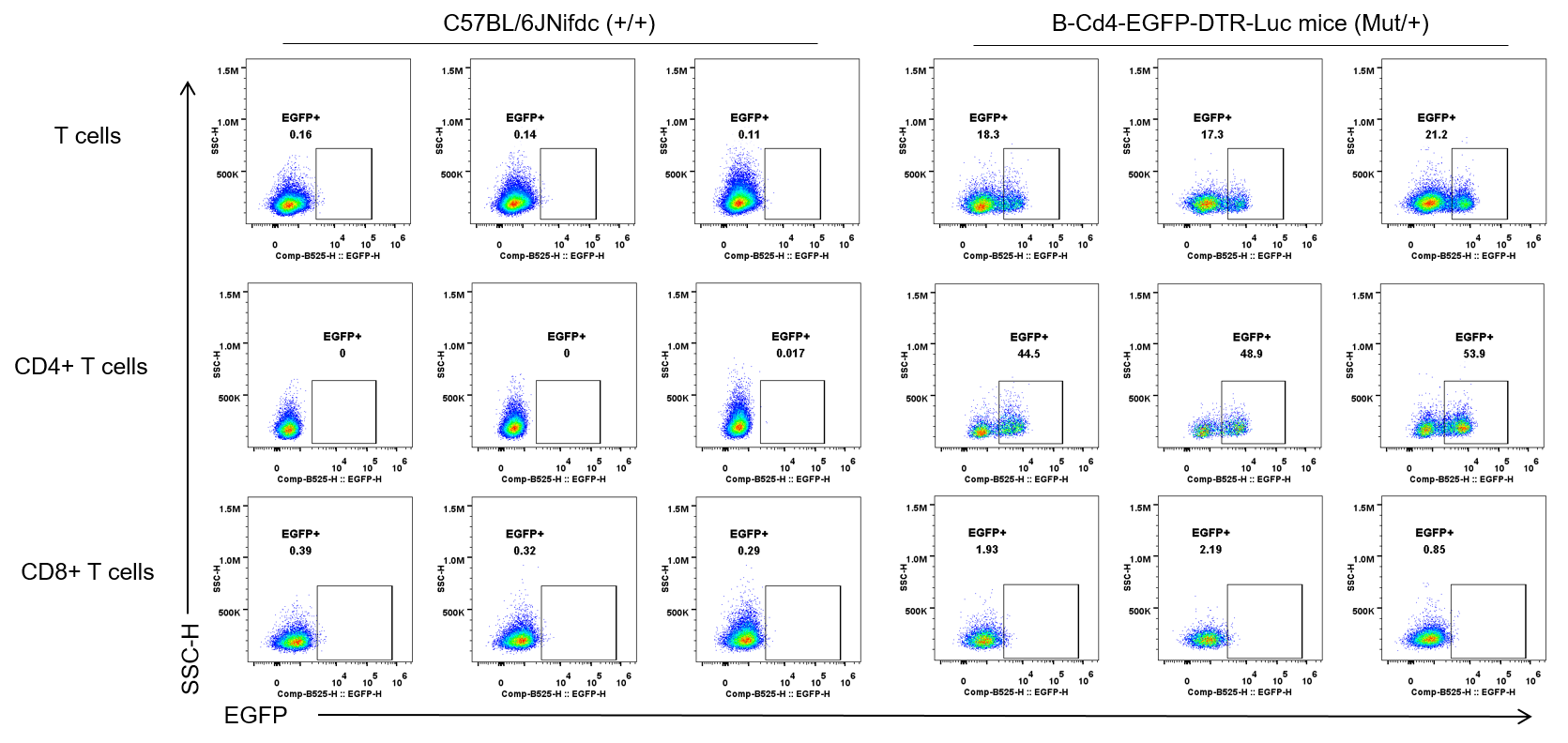
EGFP expression analysis in B-Cd4-EGFP-DTR-Luc mice by flow cytometry. Blood cells were collected from wild-type C57BL/6JNifdc mice (+/+) and heterozygous B-Cd4-EGFP-DTR-Luc mice (Mut/+)(female, 7 week-old, n=3), analyzed by flow cytometry. EGFP was detectable in T cells and CD4+ T cells from heterozygous B-Cd4-EGFP-DTR-Luc mice.
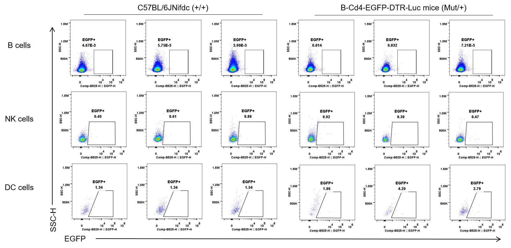
EGFP expression analysis in B-Cd4-EGFP-DTR-Luc mice by flow cytometry. Blood cells were collected from wild-type C57BL/6JNifdc mice (+/+) and heterozygous B-Cd4-EGFP-DTR-Luc mice (Mut/+)(female, 7 week-old, n=3), analyzed by flow cytometry. EGFP was not detectable in B cells, NKs and DCs from heterozygous B-Cd4-EGFP-DTR-Luc mice.
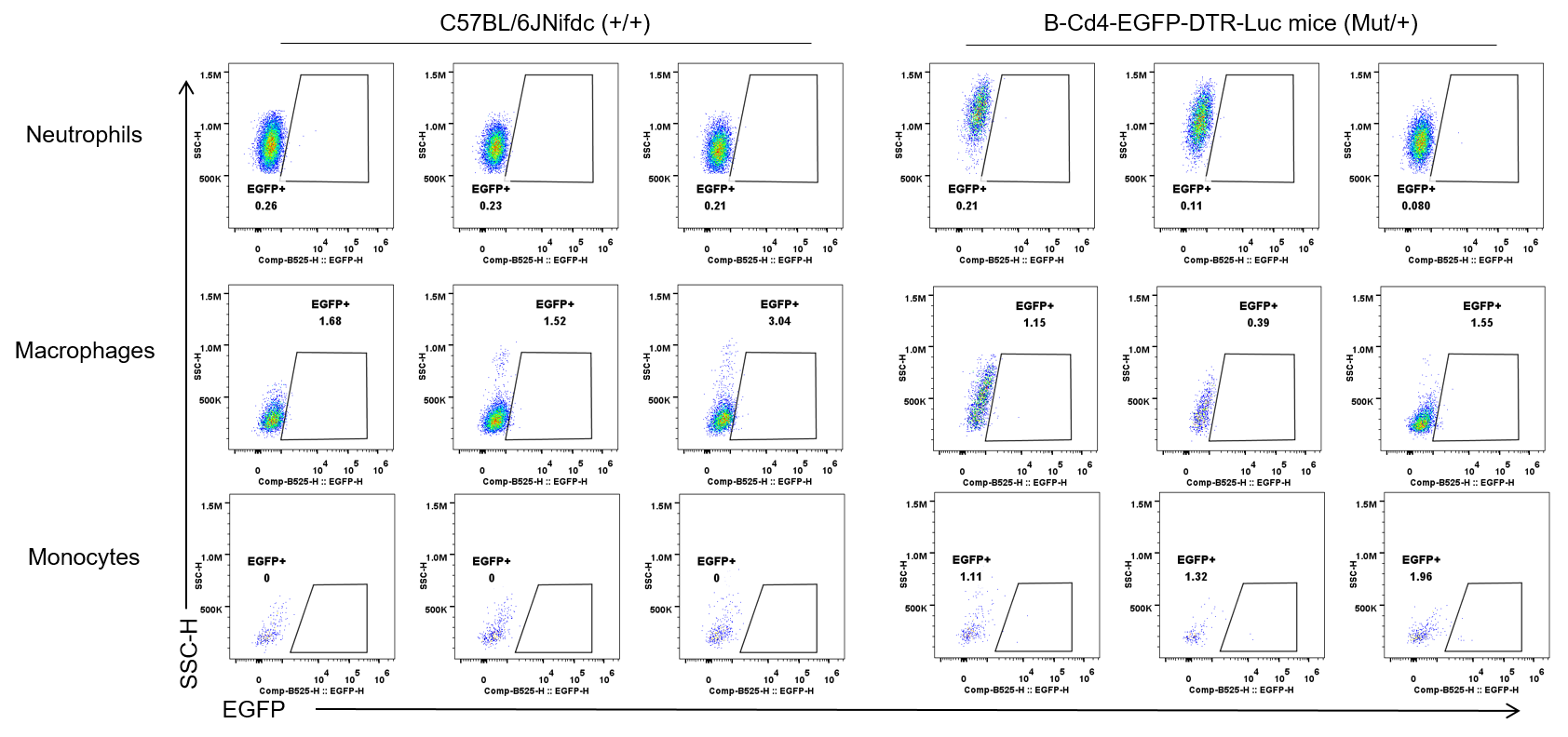
EGFP expression analysis in B-Cd4-EGFP-DTR-Luc mice by flow cytometry. Blood cells were collected from wild-type C57BL/6JNifdc mice (+/+) and heterozygous B-Cd4-EGFP-DTR-Luc mice (Mut/+)(female, 7 week-old, n=3), analyzed by flow cytometry. EGFP was not detectable in neutrophils, macrophages, and monocytes from heterozygous B-Cd4-EGFP-DTR-Luc mice.
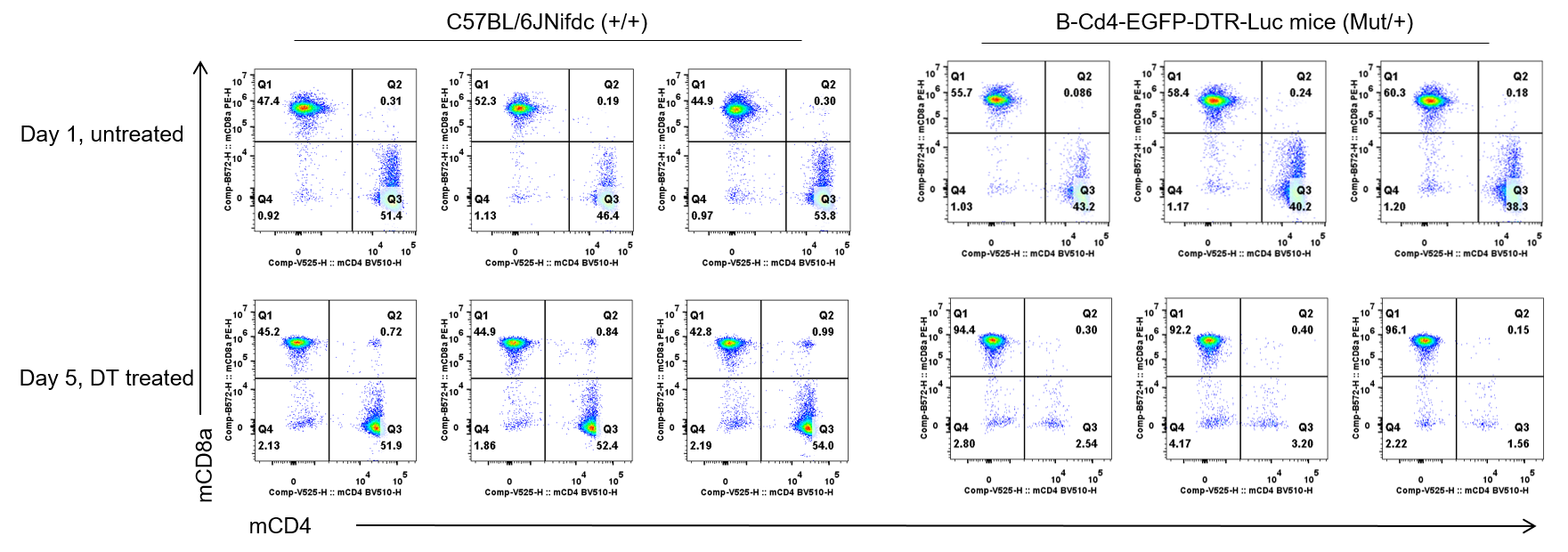
CD4+ T cells depletion analysis in B-Cd4-EGFP-DTR-Luc mice by flow cytometry. Blood cells were collected from wild-type C57BL/6JNifdc mice (+/+) and heterozygous B-Cd4-EGFP-DTR-Luc mice (Mut/+)(female, 7 week-old, n=3) injected with DT (50 ng per g body weight) for four consecutive days. Flow cytometry analysis of mouse CD4+ T cell depletion. Intraperitoneal injection of diphtheria toxin (DT) resulted in near-complete depletion of CD4+ T cells in the blood of heterozygous B-Cd4-EGFP-DTR-Luc mice, while no change was observed in wild-type (WT) mice.
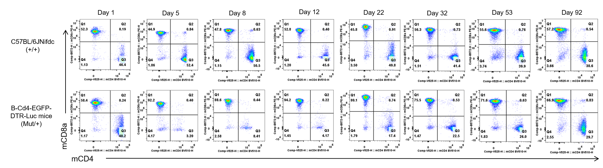
Recovery of CD4+ T cells after DT withdrawal analysis in B-Cd4-EGFP-DTR-Luc mice by flow cytometry. C57BL/6JNifdc mice (+/+) and heterozygous B-Cd4-EGFP-DTR-Luc mice (Mut/+) underwent a baseline blood collection on day 1, followed by four consecutive days of intraperitoneal injection of diphtheria toxin (DT) at a dose of 50 ng per gram of body weight. Peripheral blood samples were subsequently collected and analyzed by flow cytometry at the indicated time points (days 5, 8, 12, 22, 32, 53, and 92) to monitor CD4+ T cell dynamics. Intraperitoneal injection of diphtheria DT resulted in near-complete depletion of CD4+ T cells in the blood of heterozygous B-Cd4-EGFP-DTR-Luc mice, while no change was observed in wild-type mice. CD4+ T cell counts in blood of heterozygous B-Cd4-EGFP-DTR-Luc mice returned to near-baseline levels 87 days after DT withdrawal.
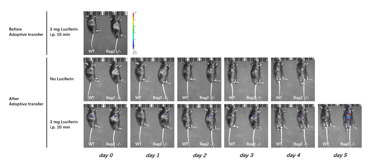
In vivo proliferation dynamics of adoptively transferred primary CD4+ T cells in syngeneic B16F10 tumor models with distinct immune niches. Wild-type (WT) C57BL/6 and Rag2 knockout (KO) mice bearing established subcutaneous B16F10 tumors received an intra-tumoral injection of 5E6 ex vivo activated primary cells isolated from a homozygous B-Cd4-EGFP-DTR-Luc mice (Mut/Mut). T cells were tracked longitudinally via bioluminescence imaging (BLI) for five consecutive days following transfer. The BLI signal rapidly declined in WT mice, indicating poor engraftment and survival of transferred cells. In contrast, Rag2 KO mice, which lack endogenous CD4+ T cells, supported signal increase, demonstrating successful engraftment and robust in vivo proliferation of the adoptively transferred CD4+ T cells. These results demonstrate that primary T cells from B-Cd4-EGFP-DTR-Luc mice are a suitable tool for adoptive transfer experiments, enabling in situ tracking of T cell proliferation in vivo.
The experimental data shown here were kindly shared by our client.