
C57BL/6-Erbb2tm1(ERBB2)Bcgen/Bcgen • 110812
| Product name | B-hHER2 mice |
|---|---|
| Catalog number | 110812 |
| Strain name | C57BL/6-Erbb2tm1(ERBB2)Bcgen/Bcgen |
| Strain background | C57BL/6 |
| NCBI gene ID | 2064 (Human) |
| Aliases | NEU; NGL; HER2; TKR1; CD340; HER-2; VSCN2; MLN 19; MLN-19; c-ERB2; c-ERB-2; HER-2/neu; p185(erbB2) |
在此页面上
Key Advantages
Validation
Application
The exons 2–17 of the mouse Erbb2 gene that encode the extracellular and transmembrane domains were replaced with human ERBB2 exons 2–17 in B-hHER2 mice.
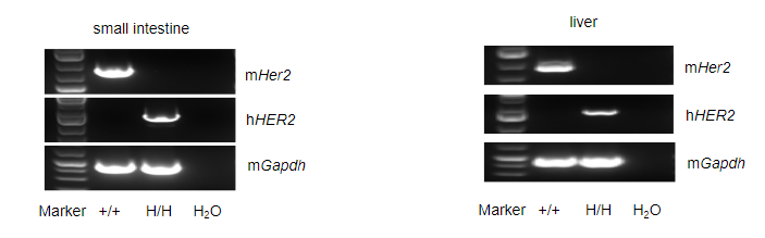
Species-specific analysis of HER2 gene expression in wild-type (WT) mice and HER2 humanized mice (B-hHER2 mice) was performed by RT-PCR. Mouse Her2 mRNA was detectable only in the small intestine and liver of WT mice (+/+). Human HER2 mRNA was detectable exclusively in homozygous HER2 humanized mice (H/H) but not in WT mice (+/+).
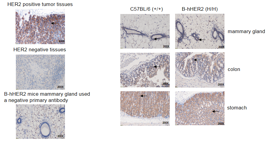
Immunohistochemical (IHC) analysis of HER2 expression was performed in homozygous HER2 humanized mice. Mammary gland, colon, and stomach tissues were collected from WT and homozygous HER2 humanized mice (H/H) and analyzed using an anti-HER2 antibody. HER2 was detectable in both WT and homozygous HER2 humanized mice due to antibody cross-reactivity. Arrows indicate tissue cells with positive HER2 staining (brown).
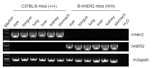
Species-specific analysis of HER2 gene expression in WT mice and HER2 humanized mice was conducted by RT-PCR. Mouse Her2 mRNA was detectable only in the eye, tongue, lung, liver, kidney, and stomach of WT mice (+/+). Human HER2 mRNA was detectable only in homozygous HER2 humanized mice (H/H) but not in WT mice (+/+).

Immunohistochemical (IHC) analysis of HER2 protein expression was performed in WT mice and HER2 humanized mice. Thirteen major tissues were collected and analyzed using anti-mouse HER2 antibody (ab214275) and anti-human HER2 antibody (ab16662). Mouse HER2 was detected in the lung, stomach, colon, small intestine, uterus, and breast of WT mice (Figure 1). Human HER2 was detected in the lung, stomach, colon, small intestine, uterus, breast, kidney, and liver of homozygous HER2 humanized mice (Figure 2). The expression profile of HER2 in HER2 humanized mice was similar to that observed in normal human tissues, indicating that HER2 humanized mice can be used to evaluate the toxicity of HER2-targeted drugs.
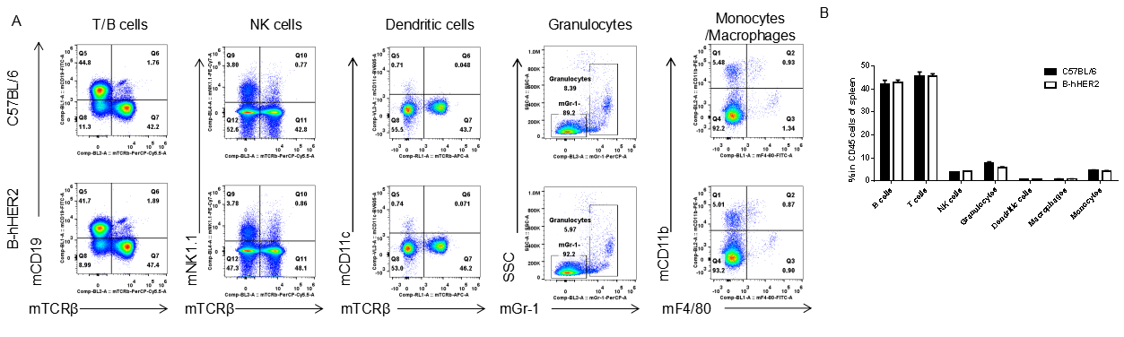
Splenocytes were isolated from female C57BL/6 and HER2 humanized mice. Flow cytometry was performed to assess leukocyte subpopulations. (A) Representative FACS plots. Single live cells were gated for CD45⁺ populations. (B) Percentages of T cells, B cells, NK cells, dendritic cells, granulocytes, monocytes, and macrophages in homozygous HER2 humanized mice were similar to those in C57BL/6 mice. This demonstrates that replacement of mouse Her2 with human HER2 does not affect leukocyte development or distribution in the spleen. N=3, 9 weeks old. Data are presented as mean ± SEM.
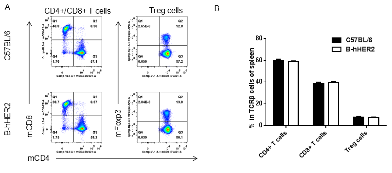
Splenocytes from female C57BL/6 and HER2 humanized mice were analyzed by flow cytometry. (A) Representative FACS plots of CD45⁺CD3⁺ T cells. (B) Percentages of CD8⁺ T cells, CD4⁺ T cells, and Tregs in homozygous HER2 humanized mice were comparable to those in C57BL/6 mice, indicating unaffected T cell development. N=3, 9 weeks old. Data are presented as mean ± SEM.
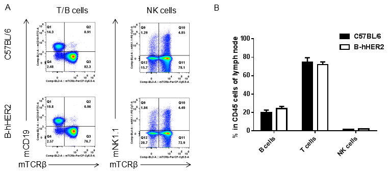
Leukocytes isolated from lymph nodes of female C57BL/6 and HER2 humanized mice were analyzed by FACS. (A) Representative plots gated on CD45⁺ cells. (B) Percentages of T cells, B cells, and NK cells were similar between HER2 humanized mice and C57BL/6 controls. N=3, 9 weeks old. Data are presented as mean ± SEM.
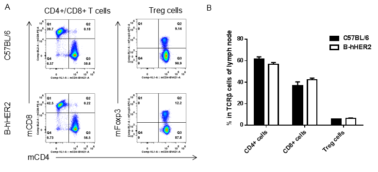
Flow cytometry analysis of lymph node T cells from female C57BL/6 and HER2 humanized mice. (A) Representative CD45⁺CD3⁺ gating strategy. (B) Percentages of CD8⁺ T cells, CD4⁺ T cells, and Tregs were comparable between groups. N=3, 9 weeks old. Data are presented as mean ± SEM.
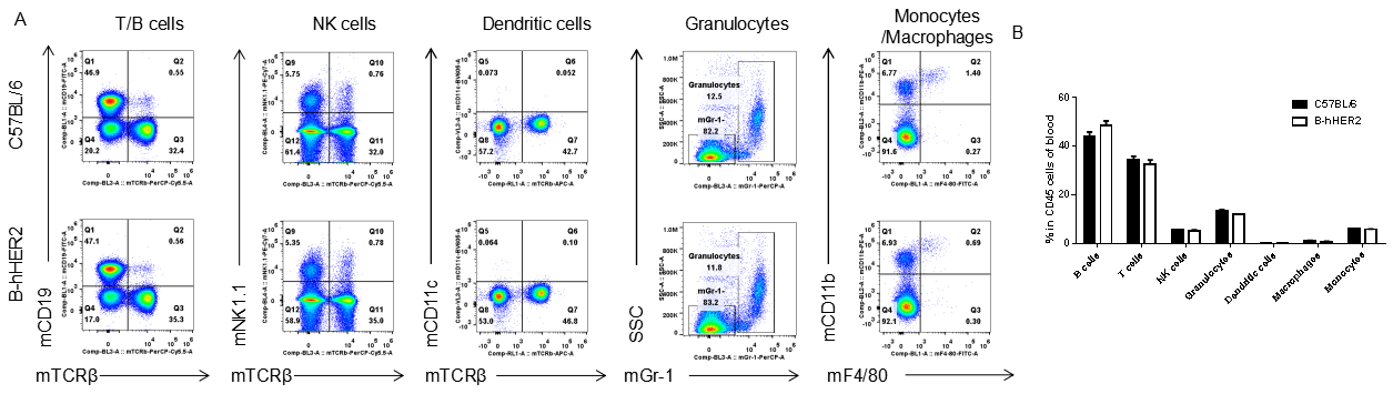
Blood leukocytes from female C57BL/6 and HER2 humanized mice were analyzed by flow cytometry. (A) Representative FACS plots gated on CD45⁺ cells. (B) Percentages of major leukocyte populations were similar between HER2 humanized mice and C57BL/6 mice. N=3, 9 weeks old. Data are presented as mean ± SEM.
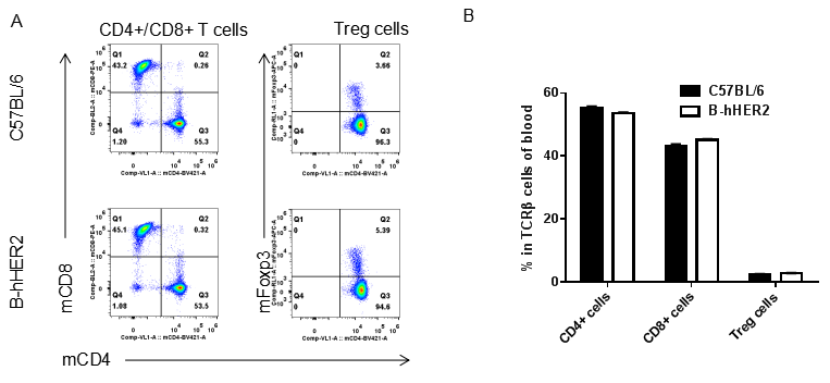
Flow cytometric analysis of blood T cell subsets from female C57BL/6 and HER2 humanized mice. (A) CD45⁺CD3⁺ gating strategy. (B) Percentages of CD8⁺ T cells, CD4⁺ T cells, and Tregs were comparable between groups. N=3, 9 weeks old. Data are presented as mean ± SEM.

Antitumor activity of anti-human HER2 ADC DS8201 (purchased from AstraZeneca/Daiichi Sankyo) in B-hHER2 mice. (A) Anti-human HER2 ADC inhibited B-hHER2 MC38 plus tumor growth in B-hHER2 mice. Murine colon cancer B-hHER2 MC38 plus cells were subcutaneously implanted into homozygous B-hHER2 mice (female, 10 weeks-old, n=6). Mice were grouped when tumor volume reached approximately 100 mm3, at which time they were intravenously injected with anti-human HER2 ADC DS8201 (purchased from AstraZeneca/Daiichi Sankyo) indicated in panel. (B) Body weight changes during treatment. As shown in panel A, anti-human HER2 ADC DS8201 (purchased from AstraZeneca/Daiichi Sankyo) was efficacious in controlling tumor growth in B-hHER2 mice, demonstrating that the B-hHER2 mice provide a powerful preclinical model for in vivo evaluation of anti-human HER2 ADC. Values are expressed as mean ± SEM.

Antitumor activity of anti-human HER2 ADC DS8201 (purchased from AstraZeneca/Daiichi Sankyo) in B-hHER2 mice. Murine colon cancer B-hHER2 MC38 plus cells were subcutaneously implanted into homozygous B-hHER2 mice (female, 10 weeks-old, n=6). Mice were grouped when tumor volume reached approximately 100 mm3, at which time they were intravenously injected with anti-human HER2 ADC DS8201 (purchased from AstraZeneca/Daiichi Sankyo) indicated in panel.

Antitumor activity of anti-human HER2 antibody (Trastuzumab analog-MMAE, in-house) in B-hHER2 mice. (A) Anti-human HER2 antibody inhibited B-hHER2 MC38 plus tumor growth in B-hHER2 mice. Murine colon cancer B-hHER2 MC38 plus cells were subcutaneously implanted into homozygous B-hHER2 mice (female, 7-8-week-old, n=6). Mice were grouped when tumor volume reached approximately 100 mm3, at which time they were intravenously injected with anti-human HER2 ADC Trastuzumab analog-MMAE (in-house) indicated in panel. (B) Body weight changes during treatment. As shown in panel A, anti-human HER2 ADC Trastuzumab analog-MMAE (in-house) was efficacious in controlling tumor growth in B-hHER2 mice in a dose-dependent manner, demonstrating that the B-hHER2 mice provide a powerful preclinical model for in vivo evaluation of anti-human HER2 antibodies. Values are expressed as mean ± SEM.
The overage of this tumor model is 40%.

Antitumor activity of anti-human HER2 antibody (Trastuzumab analog-MMAE, in-house) in B-hHER2 mice. (A) Anti-human HER2 antibody inhibited B-hHER2 MC38 plus tumor growth in B-hHER2 mice. Murine colon cancer B-hHER2 MC38 plus cells were subcutaneously implanted into homozygous B-hHER2 mice (female, 7-8-week-old, n=6). Mice were grouped when tumor volume reached approximately 100 mm3, at which time they were intravenously injected with anti-human HER2 ADC Trastuzumab analog-MMAE (in-house) indicated in panel.

Antitumor activity of anti-human HER2 antibody (DS8201, purchased from Daiichi Sankyo) in B-hHER2 mice. (A) Anti-human HER2 antibody inhibited B-hHER2 MC38 plus tumor growth in B-hHER2 mice. Murine colon cancer B-hHER2 MC38 plus cells were subcutaneously implanted into homozygous B-hHER2 mice (female, 7-8-week-old, n=6). Mice were grouped when tumor volume reached approximately 100 mm3, at which time they were intravenously injected with anti-human HER2 ADC DS8201 (purchased from AstraZeneca/Daiichi Sankyo) indicated in panel. (B) Body weight changes during treatment. As shown in panel A, anti-human HER2 ADC DS8201 (purchased from Daiichi Sankyo) was efficacious in controlling tumor growth in B-hHER2 mice in a dose-dependent manner, demonstrating that the B-hHER2 mice provide a powerful preclinical model for in vivo evaluation of anti-human HER2 antibodies. Values are expressed as mean ± SEM.
The overage of this tumor model is 40%.

Antitumor activity of anti-human HER2 antibody (DS8201, purchased from Daiichi Sankyo) in B-hHER2 mice. (A) Anti-human HER2 antibody inhibited B-hHER2 MC38 plus tumor growth in B-hHER2 mice. Murine colon cancer B-hHER2 MC38 plus cells were subcutaneously implanted into homozygous B-hHER2 mice (female, 7-8-week-old, n=6). Mice were grouped when tumor volume reached approximately 100 mm3, at which time they were intravenously injected with anti-human HER2 ADC DS8201 (purchased from AstraZeneca/Daiichi Sankyo) indicated in panel.
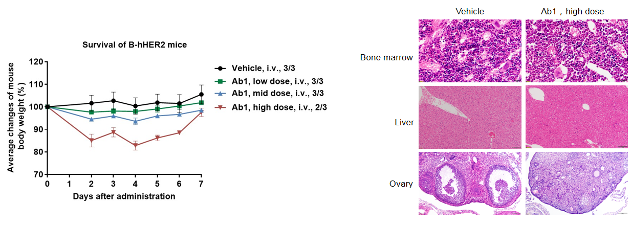
Different doses of anti-human HER2 antibody (Ab1) were administered to HER2 humanized mice via a single intravenous injection. Body weight decreased in a dose-dependent manner, and one mouse died in the high-dose group. Histopathological analysis revealed no significant liver abnormalities; however, ovaries lacked immature follicles, myeloid cells increased in bone marrow, and red blood cell counts decreased. These results suggest that HER2 humanized mice can be used to assess the toxicity of anti-human HER2 antibodies. Data are presented as mean ± SEM. This data is part of a collaboration with a partner.
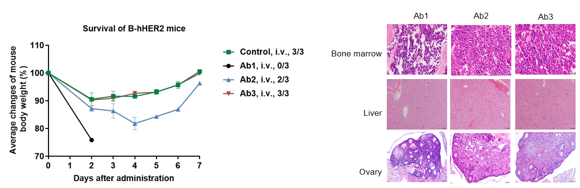
Different anti-human HER2 antibodies were administered to HER2 humanized mice via single intravenous injection. Weight loss varied among treatment groups. In the Ab1 group, all mice were euthanized due to rapid weight loss. Mild toxicity was observed in bone marrow and ovaries by histopathological analysis. These results further support the application of HER2 humanized mice in evaluating the toxicity of anti-HER2 antibodies. Data were obtained from a partner. Data are presented as mean ± SEM.
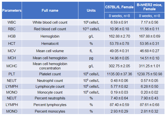
Complete blood count (CBC) analysis was performed using blood from female C57BL/6 and HER2 humanized mice. No differences were observed between groups, indicating that replacement of mouse Her2 with human HER2 does not affect blood cell composition or morphology. N=8, 8 weeks old. Data are presented as mean ± SEM.
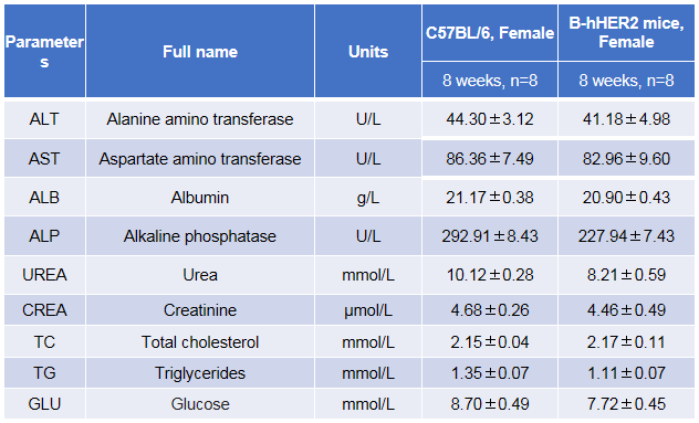
Blood chemistry analysis in female C57BL/6 and HER2 humanized mice. No differences were observed between groups, indicating normal liver function. N=8, 8 weeks old. Data are presented as mean ± SEM.
Q1: What is the main application of HER2 humanized mice (B-hHER2)?
HER2 humanized mice are primarily used for preclinical toxicity and safety evaluation of anti-HER2 antibodies and biologics, enabling assessment of on-target effects in a human-relevant in vivo setting.
Q2: How is HER2 expression validated in HER2 humanized mice (B-hHER2)?
HER2 expression in HER2 humanized mice is validated at both the mRNA and protein levels using RT-PCR and immunohistochemistry. Human HER2 expression is detected exclusively in homozygous HER2 humanized mice, with tissue distribution patterns similar to those observed in normal human tissues.
Q3: Does humanization of HER2 affect the immune system of the mice?
No. Comprehensive flow cytometry analyses demonstrate that leukocyte and T cell subpopulation distributions in spleen, lymph nodes, and peripheral blood of HER2 humanized mice are comparable to those of wild-type C57BL/6 mice, indicating preserved immune system development and homeostasis.
Q4: What types of drugs can be evaluated using HER2 humanized mice (B-hHER2)?
HER2 humanized mice can be used to evaluate the safety and toxicity of anti-HER2 monoclonal antibodies, antibody–drug conjugates (ADCs), and other HER2-targeted biologics that specifically recognize human HER2.